Almost all energy required for blood flowing through our arteries and veins is delivered by the heart. A small amount is delivered by a short-lasting contraction of the arteries at every heart beat onset; that, at least, is the proposition of the theory of arterial acceleration.
It is important that we realize the heart is a volume pump, not a pressure pump. The most important argument is that the heart delivers its energy in a pulsed fashion. During systole the heart pumps its volume into the arterial tree. Part of the volume is added to the volume already present in the arterial tree allowing cardiac energy to be transformed to potential energy generated by stretch of the arterial wall. Part of the volume pushes forward the volume already present, forcing blood out into the many capillary networks. During diastole, it is the potential energy of blood volume stored in the arterial tree that forces out blood from arterioles into capillaries.
Let us briefly imagine the situation where the wall of the arterial tree is non-compliant, as if it were made of stainless steel. Then any volume pushed in on the one side of the system would inevitably and instantaneously lead to the same volume being pushed out on the other side. Broader pipes have less resistance than smaller ones and will allow more blood to flow. Longer pipes have a higher resistance than shorter ones and will receive less blood to flow. There is no pressure gradient, since fluid cannot be compressed and no buildup of pressure can occur when it leads to instantaneous relief over all the branches.
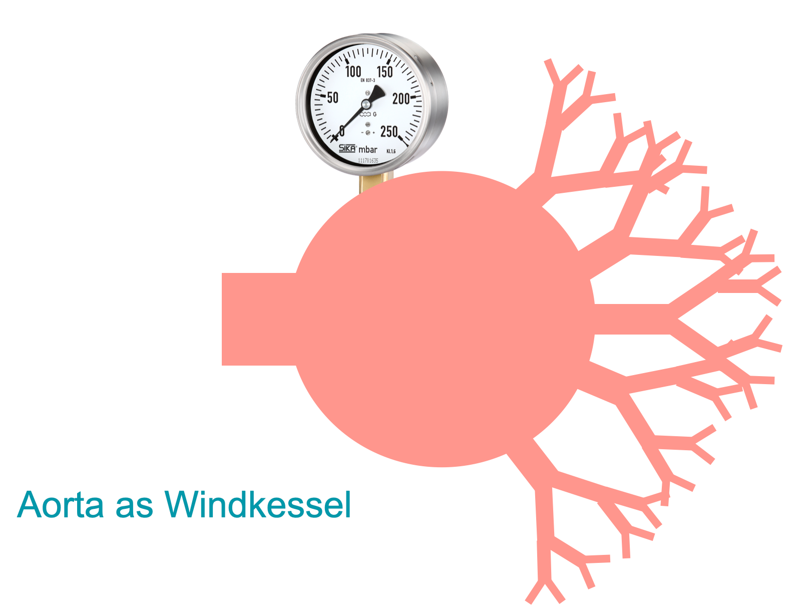
The figure above represents a caricature of the windkessel model, with a pressurized wide body capacitance and many outflow resistances to be regulated for blood supply to the different destinations. Traditionally, organs or thought to adjust their arteriolar resistance locally by means of metabolic coupling, or their blood supply is regulated by humoral or neural control. By no means can the heart preferentially direct its flow towards one or more organs, leaving other organs out. Instead, the heart forces its stroke volume into the narrow end of a funnel with an immense increase in overall cross-sectional area from proximal to distal.
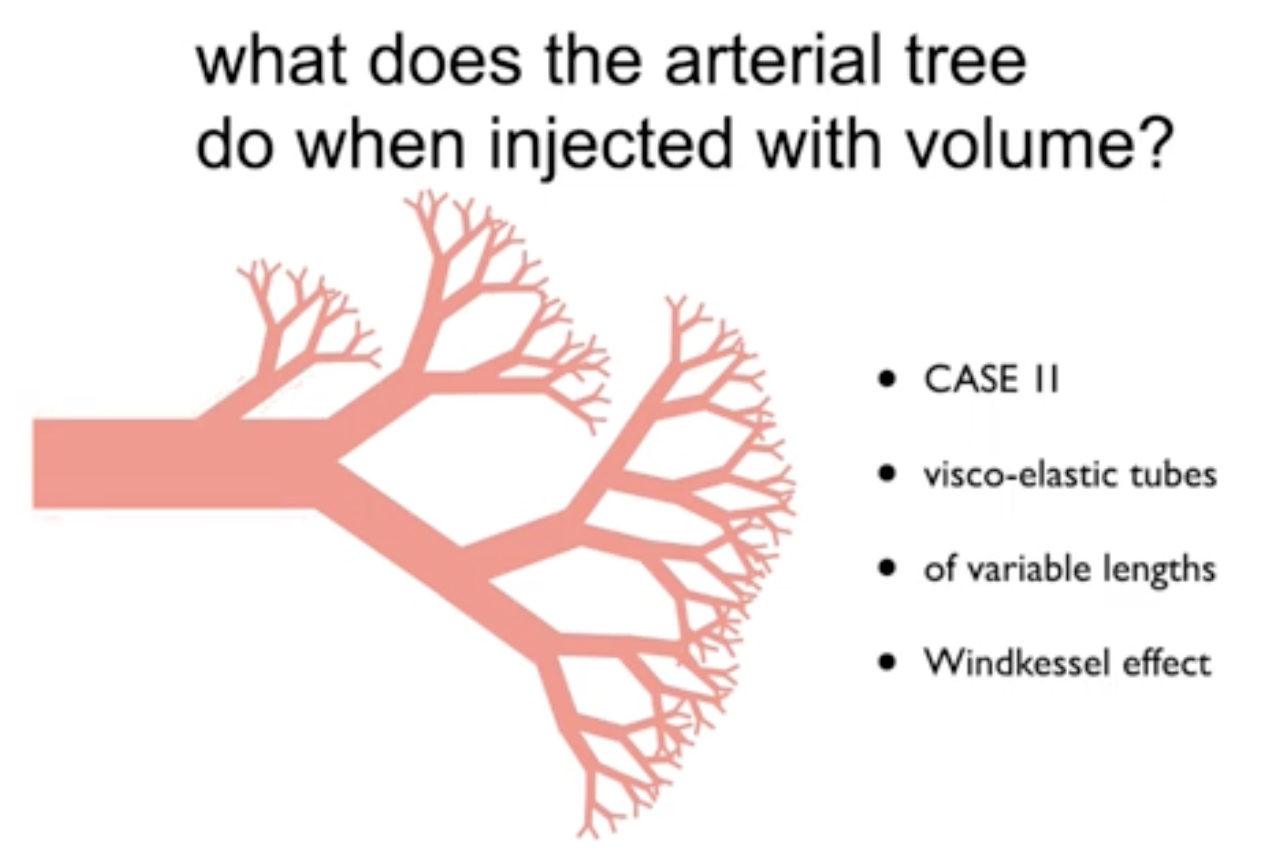
Given that the arterial resistance will greatly differ from branch to branch the system risks ill-perfusion of regions with somewhat higher resistance in favor of those with a reduced resistance or a less remote localization along the arteriolar tree. How can the arterial tree ensure that all the bodies capillary systems are at least perfused with minimal flow?

The theory of arterial acceleration proposes that the first increase in pressure in the proximal aorta triggers a myogenic response in its smooth muscle cells: a so-called stretch induced depolarization. The smooth muscle cells are electronically coupled causing this depolarization to spread from proximal to distal along the contractile layers of the arterial tree. Inside a single smooth muscle cell, this depolarization induces a Ca-spike: a brief inflow of extracellular calcium that (still to be proven) causes a slight and short-lasting contraction of the cell. Smooth muscle cells in arteries are arranged circularly and not longitudinally. Together, this wave of depolarization and the contraction within circularly arranged smooth muscle cells is thought to move along the branches of the arterial tree and bring blood into motion even in the most remote capillary systems of the body. The propagation speed of this wave (pulse wave velocity or PWV) can be calculated to increase from about 2 in the more proximal arteries to 8-10 m/s in the more distal. Its relative contribution gains in importance with respect to the pressure wave generated by heart contraction, since arterial acceleration builds upon the myogenic response in more proximal branches where the pressure wave dilutes along its trajectory: there is a great increase in overall cross-sectional area making the arterial tree a sort of funnel with the heart pumping blood into its narrow end.

Consequently, the Windkessel figure above is an over-simplification of what is really happening in the arterial tree. It leads to the common misunderstanding that a patient has only a single arterial blood pressure. Instead, it should be realized that arterial blood pressure varies with location and with time. Treatment decisions can be made on instant measurements only when the measurement location and method is standardized and when its systolic waveform is correctly analyzed for its different components as below.

It is an important observation that the two components of the systolic pressure wave vary with age. Aging takes a toll on arterial elasticity and arteries dilate and elongate when we become older. The loss of elasticity in the proximal arteries causes the well known increase in arterial stiffness. The capacitance in which the heart pumps its gradually decreasing stroke volume (aging also goes with a reduction of cardiac ejection fraction) becomes sloppy, its mechanics relying more on collagen than on elastin. More blood is required to fill the capacitance vessels, the relative contribution of stroke volume becomes less and circulation time will become larger since blood lingers longer in the arterial phase. Because of the increase in stiffness, the pressure increase by addition of stroke volume is steeper and more blood is forced out during systole. During diastole, however, due to a rapid decrease in pressure diastolic blood flow approaches zero. This causes the pulsatility index (difference between systolic and diastolic pressure or flow divided by mean pressure or flow) to increase with old age.

Arterial acceleration decreases with aging: arterial blood pressure and intracranial blood flow recordings show that the systolic waveform is Sys1 dominant in the young but becomes increasingly Sys2 dominant in the old. The dilatation of capacitance arteries, causing stretch of the smooth muscle cells, is possibly detrimental for the underlying short-lasting contraction of the myogenic response.

For a rapid interpretation of middle cerebral artery flow velocity recordings we have proposed a radar plot of age corrected Z-scores. Z-scores are calculated by dividing the difference between an actual measurement and the mean value expected for that age through the observed standard deviation. In this way any measurement can easily be weighed to what is expected for that age and the four components of the waveform (Sys1, Sys2, D560 and HR) can be compared to each other.

Above a radar plot is shown for the four components of middle cerebral artery flow velocity during hyperventilation (red), normoventilation (green) and CO2-retention (yellow).

The penetration force of Sys1 into remote capillary systems is larger than of Sys2 and diastolic blood flow since it builds upon the wave of arterial acceleration in more proximal arteries. Metabolic coupling determines whether arterioles dilate or constrict letting in more or less blood adding to the blood flow generated by arterial acceleration. Arterial acceleration also makes sure that tissue is perfused despite unfavorable conditions, such as when experiencing external pressure. A simple experiment with a blood pressure cuff positioned around the hand suggests that Sys1 flow or systolic spikes may even occur despite inflating the cuff to pressures well over 150 mmHg. Sys1 perfusion ensures that local ischemia due to, for instance, high tissue pressures will not go unnoticed to the central control centers (chemoceptors) allowing the organism as a whole to respond with an increase in blood pressure and/or respiratory activity (CNS ischemic response). Without Sys1 perfusion local ischemia may remain unnoticed as blood cannot enter into a non-perfused tissue with accumulating signs of local ischemia.
From a physiological perspective arterial acceleration is a welcome addition to the pumping activity of the heart allowing blood flow to occur into all the bodies capillary systems, including those that are most remote and under least favorable conditions.